Understanding the Structure and Function of the Human Chest
The chest is one of the most vital regions of the human body, housing essential organs that play a key role in breathing, circulation, and overall body function. Known scientifically as the thoracic region, the chest human anatomy is a complex combination of bones, muscles, and internal organs protected by the rib cage. Learning more about the parts of chest anatomy can help you understand how the body maintains life and responds to injury or disease.
Main Components of the Chest Anatomy
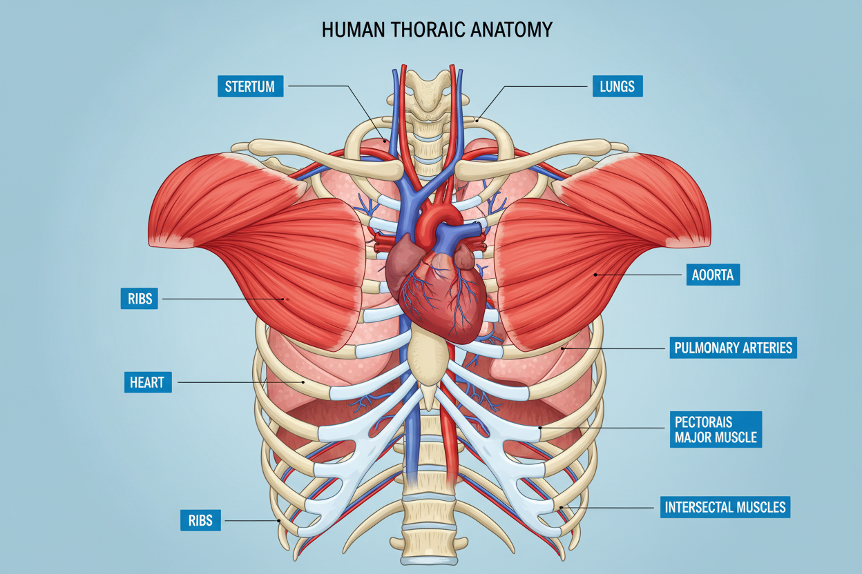
When discussing the parts of the chest anatomy, it is important to recognize the three major systems it contains: the skeletal structure, muscular components, and internal organs. Each of these systems works together seamlessly to enable vital functions such as respiration and cardiovascular circulation. The chest human anatomy is protected primarily by the thoracic cage, which comprises 12 pairs of ribs, the sternum, and thoracic vertebrae.
Skeletal Structure
The skeletal framework of the chest anatomy consists mainly of bones and cartilage. The sternum, or breastbone, lies at the center front, connecting to ribs via costal cartilage. The ribs form a semi-flexible cage around the thoracic cavity, allowing expansion and contraction during breathing. Behind the chest cavity lies the spine’s thoracic segment, supporting posture and providing anchoring points for muscles.
Muscular System
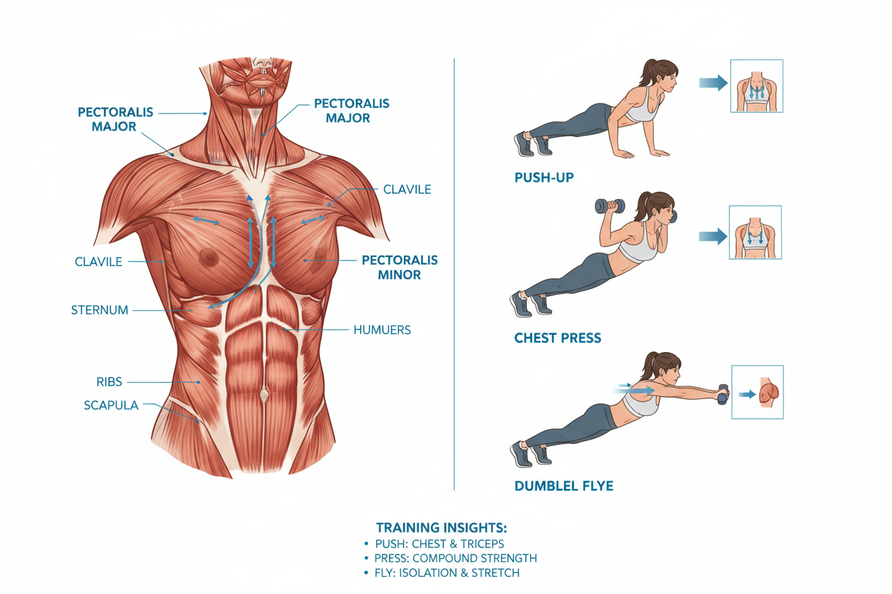
In addition to bones, the chest anatomy houses powerful muscles that facilitate breathing and upper body movement. The intercostal muscles are situated between the ribs, aiding in the expansion and contraction of the chest during inhalation and exhalation. The diaphragm, a dome-shaped muscle beneath the lungs, is another critical component. It contracts and flattens during inhalation, increasing the chest cavity's volume and drawing air into the lungs.
Internal Organs
Within the chest human anatomy lie the lungs and heart — two of the body’s most important organs. The lungs are responsible for oxygen intake and the exchange of carbon dioxide, while the heart pumps blood throughout the body. The esophagus and sections of major blood vessels such as the aorta and vena cava also run through this region. These parts of chest anatomy work closely in maintaining overall body health.
Functionality and Interaction of Chest Components
The chest is an integrated system where skeletal, muscular, and organ functions influence each other. For example, when you inhale, the ribs move outward and upward, aided by the intercostal muscles, while the diaphragm lowers to create space for lung expansion. Simultaneously, the heart adjusts blood flow to align with increased oxygen intake. In conditions like asthma or heart disease, disruptions in one part of the chest anatomy can affect the others, leading to symptoms such as shortness of breath or chest pain.
Clinical Relevance
Understanding the parts of chest anatomy is crucial for identifying potential health problems. Medical professionals often use physical exams, imaging techniques, and auscultation to assess chest health. For example, chest X-rays can reveal rib fractures, lung infections, or enlarged heart sizes. A clear knowledge of the chest human anatomy helps in diagnosing conditions like pneumonia, pulmonary embolism, or musculoskeletal injuries.
Common Conditions Affecting the Chest
Lung diseases such as chronic obstructive pulmonary disease (COPD) and asthma directly impact breathing mechanics. Heart conditions like myocardial infarction originate in the chest cavity and can be life-threatening. Musculoskeletal injuries, such as rib fractures, may cause significant pain and restrict movement. In all these conditions, proper medical evaluation of the chest anatomy is necessary for effective treatment.
Personal Experience with Understanding Chest Anatomy
During my time volunteering in a community health program, I met patients who had little knowledge of their own chest anatomy and how it related to their symptoms. After explaining the function of ribs, muscles, and the heart, many became better at describing their discomfort to physicians, leading to more accurate diagnoses. This experience reinforced that knowing the parts of chest anatomy is not just a medical necessity but also empowers individuals to take control of their health.
Conclusion
The chest human anatomy is a remarkable combination of protective skeletal structures, powerful muscles, and critical organs. Each part of the chest anatomy plays an essential role in sustaining life. Whether you are studying anatomy for professional purposes or for personal awareness, understanding the chest's components and their interaction provides valuable insight into overall health and disease prevention.
