Gluteus Maximus Cadaver Anatomy: Structural Borders, Muscle Layers, and Functional Floor Staging
Gross Anatomy of the Gluteus Maximus: Insights from Cadaveric Observation
In anatomical dissection, the gluteus maximus cadaver specimen immediately stands out due to its sheer scale and distinctive coarse texture. Unlike the tightly bundled, parallel fibers found in many limb flexors, the superficial gluteal layer displays large, heavy fascicles bound by substantial sheets of fibroelastic tissue. When the overlying skin and subcutaneous adipose tissue are carefully reflected, the muscle reveals a broad, quadrate-to-rhomboid shape that dominates the posterior pelvic basin.
Anatomists and biomechanics observers frequently note that the fascicular arrangement of the gluteal muscles cadaver complex is organized diagonally rather than vertically. The muscle fibers run downward and outward across the buttock region. This structural architecture reflects its primary functional role: generating massive extension forces while stabilizing the pelvis against forward tipping of the torso. Examining an intact dissection highlights how the dense epimysium forms structural compartmentalization, preserving muscle integrity across diverse hip angles and weight-bearing tasks.
Anatomical Borders and Attachments of the Gluteus Maximus
Understanding the clear borders of the gluteus maximus provides essential context for how tension distributes across the pelvic girdle and femur during multi-joint compound movements. In a typical dissection, the borders are traced as follows:
- Superior Border: A thin, oblique boundary running from the iliac crest toward the greater trochanter, resting closely over the deep investing fascia of the gluteus medius.
- Inferior Border: A thick, free margin that forms the physical fullness of the buttock and crosses the horizontal gluteal fold obliquely as it travels toward the femoral shaft.
- Medial Border: The extensive proximal origin anchoring along the posterior gluteal line of the ilium, the posterior surface of the lower sacrum, the lateral border of the coccyx, and the fibrous tissue of the sacrotuberous ligament.
- Lateral Border: The distal destination, separating into two distinct structural insertions that dictate its force transfer.
The distal insertion observed in a gluteus maximus cadaver dissection reveals a functional bifurcation. The larger superficial portion—representing the majority of the upper muscle belly and the superficial half of the lower section—merges directly into the iliotibial band of the fascia lata. The deeper portion of the lower muscle fibers anchors securely into the gluteal tuberosity on the posterior surface of the proximal femur, between the vastus lateralis and the adductor magnus. This dual insertion provides structural anchoring across both the femur and the lateral knee complex.
Deep Layer Relationships: Gluteus Medius and the Pelvic Stabilizers
Reflecting the gluteus maximus laterally along its origin uncovers a distinct anatomical plane housing the middle and deep gluteal compartments. The gluteus medius cadaver layer sits directly beneath the anterosuperior portion of the maximus, presenting a fan-shaped arrangement that originates along the outer iliac blade between the anterior and posterior gluteal lines. The medius converges downward into a stout, flattened tendon that anchors on the lateral surface of the greater trochanter.
Beneath the lower half of the gluteus maximus sits an array of deep lateral rotators and vital neurovascular pathways. Dissectors observe the pear-shaped piriformis crossing the greater sciatic foramen, accompanied by the superior and inferior gemelli, the obturator internus tendon, and the quadratus femoris. Emerging beneath the piriformis is the substantial trunk of the sciatic nerve. In our editorial view, observing these layered structures side by side makes it immediately obvious that superficial hip power depends on the structural stability provided by these deeper pelvic stabilizers.
Translating Cadaveric Architecture into Biomechanical Vector Training
The diagonal fiber trajectory visible in human gross anatomy explains why simple planar training rarely maximizes muscle fiber recruitment across the entire gluteal complex. Because the fibers descend at an outward angle from the axial skeleton toward the appendicular skeleton, the muscle acts as a multi-vector engine. Its primary action is hip extension, but the orientation of the upper fibers enables hip abduction, while the lower fibers strongly contribute to external rotation.
To align training with this fiber geometry, loaded movements benefit from combining sagittal extension with rotational or lateral demands. When setting up compound hip exercises, reviewing a comprehensive bum muscles guide helps clarify how alternating between hip hinges, hip thrusts, and multi-directional lunges engages both the deep rotators and the superficial prime movers effectively.
Ground Reaction Forces and Floor Staging for Glute Activation
Because the gluteus maximus attaches through the iliotibial tract and the gluteal tuberosity, muscular tension generated at the hip must transfer down the kinetic chain into the ground. When executing heavy barbell hip thrusts, deadlifts, or deep squats, foot placement and subfloor firmness directly influence force transmission.
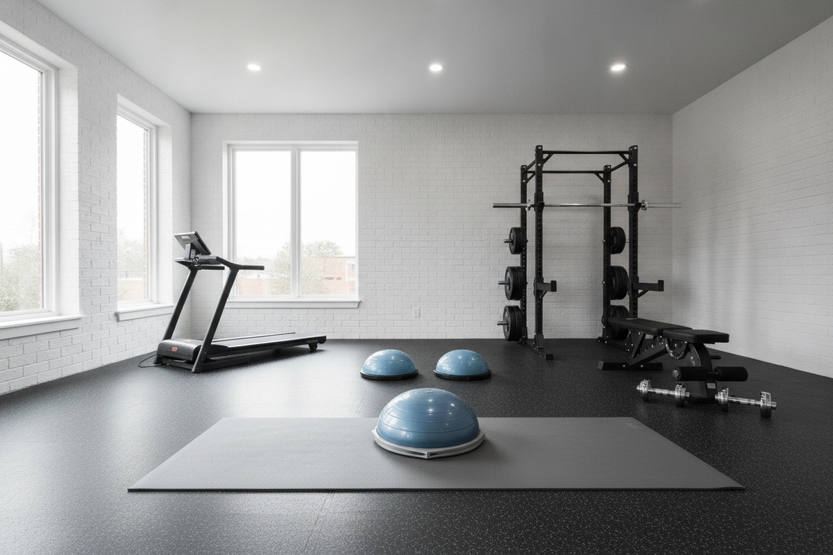
Using a squishy, overly soft surface can cause micro-instability at the foot and ankle, leading to compensations through the knee and hip that detract from clean gluteal recruitment. Conversely, a firm, high-density exercise mat or dense rubber flooring provides an unyielding base that allows lifters to drive through the heels and maintain external rotational torque. Staging your stance on an unyielding, high-traction foundation creates the structural support needed when exploring different foot widths, as detailed in our squat muscles guide.
Equipment Placement and Surface Protection for Lower-Body Training
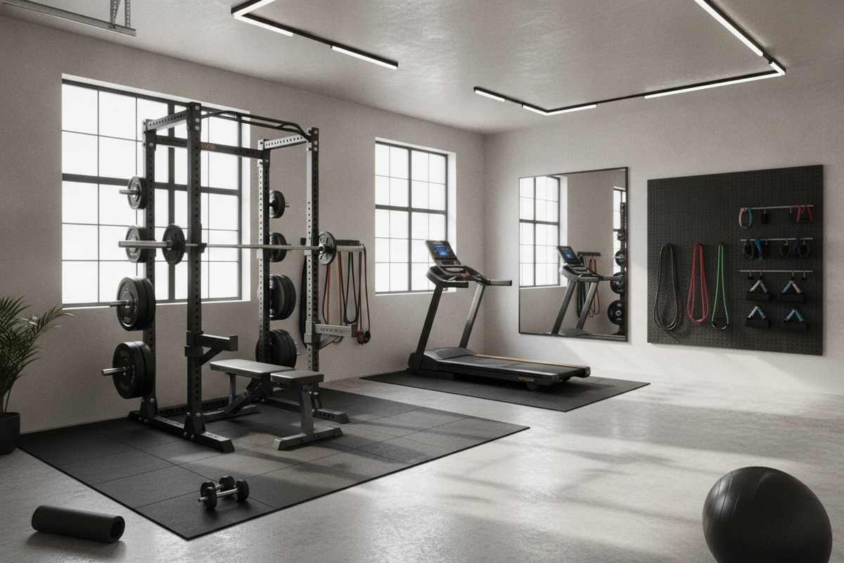
Executing high-load lower-body training requires thoughtful layout and surface considerations to preserve home gym floors and maintain lifting stability. High-force hip movements, such as weighted thrusts and loaded split squats, concentrate significant downward and lateral pressure on specific zones of your training space.
- Bench Anchoring: When setting up a bench for hip thrusts, place the bench feet directly on non-slip rubber mats. This prevents the bench from sliding backward under horizontal driving forces and stops the bench frame from denting wood or tile subfloors.
- Foot Traction Zones: Ensure the area directly beneath the feet offers a textured, high-grip finish. A slip-resistant rubber surface enables athletes to maintain an active arch and lateral foot pressure without footwear shifting.
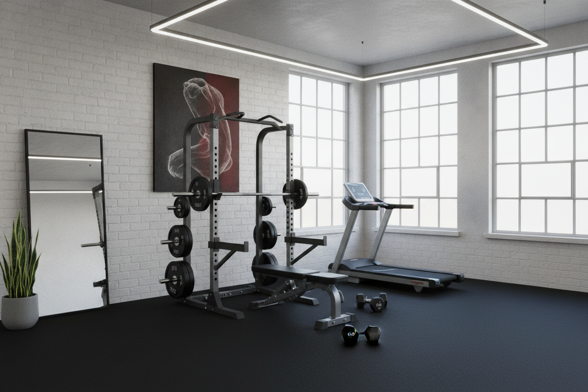
- Barbell Impact Protection: Lowering heavy bars during Romanian deadlifts or resting loaded barbells between hip thrust sets can damage concrete or finished flooring. High-density rubber mats distribute point loads over a larger surface area, mitigating impact wear on equipment and subfloors alike.
- Clear Clearance Pathways: Keep adjacent floor space free from stray weight plates, resistance bands, or loose accessories, allowing unobstructed range of motion during lateral lunges and dynamic warm-up drills.
By pairing insights from cadaveric anatomy with stable flooring choices and deliberate space staging, lifters can perform demanding lower-body strength work with confidence and solid mechanical support.